
Figure 2. Off-response currents in oocytes expressing PKD2L1 with or without PKD1L3.
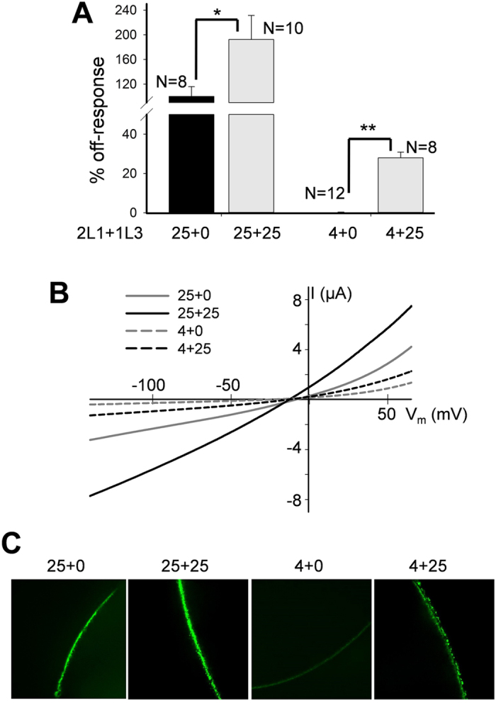
(A) Averaged and normalized off-response currents elicited by extracellular pH 3.0 from oocytes injected with 4 or 25 ng of PKD2L1 (2L1) mRNA alone or co-injected with 25 ng of PKD1L3 (1L3) mRNA, and voltage clamped at −50 mV. Shown are off-response currents averaged from different numbers of oocyte, as indicated. *p = 0.05 and **p = 0.004, unpaired t-test. (B) Representative off-response I-V curves obtained using a ramp protocol under the same conditions as in panel A. (C) Representative immunofluorescence data showing the PM intensity of PKD2L1 protein in oocytes injected with different amounts of mRNAs, as indicated.