
Figure 7. Role of extracellular Ca2+ ions in the PKD2L1 off-response.
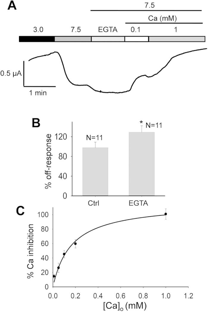
(A) Representative current recorded at −50 mV in a PKD2L1-expressing oocyte under various extracellular conditions, as indicated. (B) Based on experiments described in panel A, averaged data to show the effect of extracellular EGTA (1 mM). *p = 0.04, unpaired t-test. (C) Dose-dependence of the relative Ca2+ inhibition of the off-response current. Data were averaged from 11 oocytes and the curve is a fit of the data to Equation 1, with Ki value of 167 ± 33 μM.