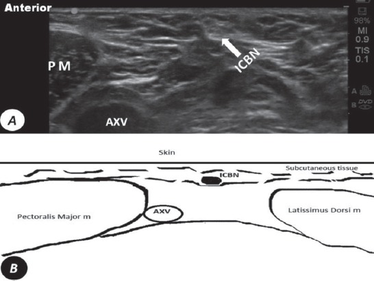
Figure 2.

A) Ultrasound short axis view at the base of the axillary area shows the intercostobrachial nerve (ICBN) as an oval shaped hyper-echoic structure posterior to lateral border of the pectoralis major muscle, superior, and posterior to the axillary vein (AXV). B) Diagram of short axis view at the base of the axillary area. PM - pectoralis major muscle
