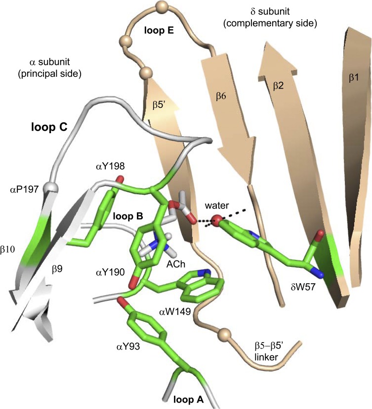
Figure 1.
The ligand-binding site of an acetylcholine binding protein. The agonist-binding sites are at subunit interfaces; the principal side (α subunit in AChRs) is white, and the complementary side (δ, ε, or γ subunit) is tan. The structure is Lymnaea stagnalis (Protein Data Bank accession no. 3WIP; Olsen et al., 2014), and residue numbers are mouse endplate AChRs. Green, aromatic core; tan spheres, αC atoms of γ-subunit substitutions (see Fig. 6); red sphere, structural water; dashed lines, H bonds.

