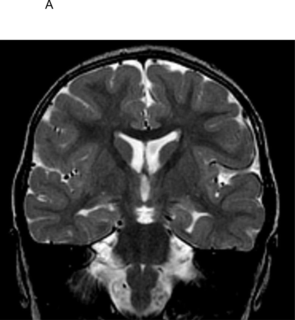
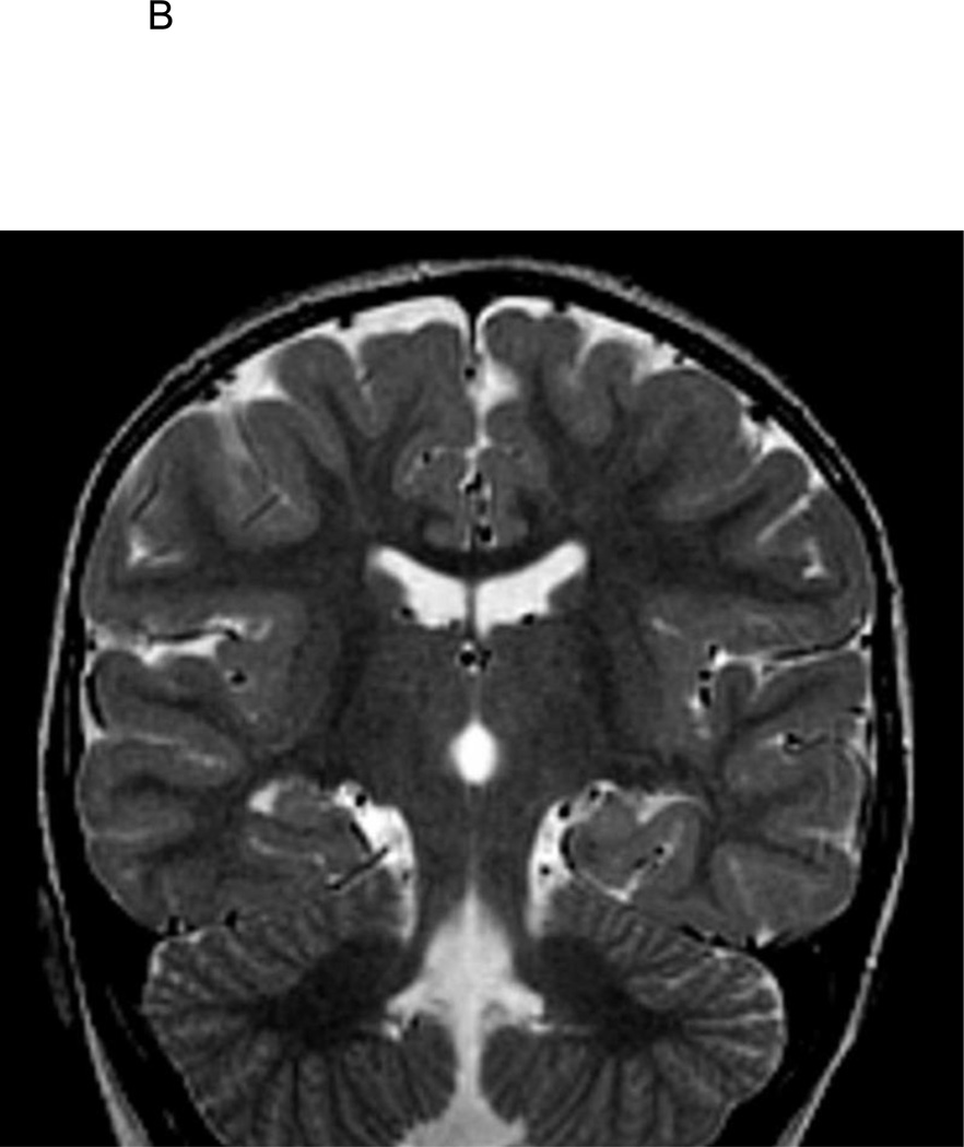
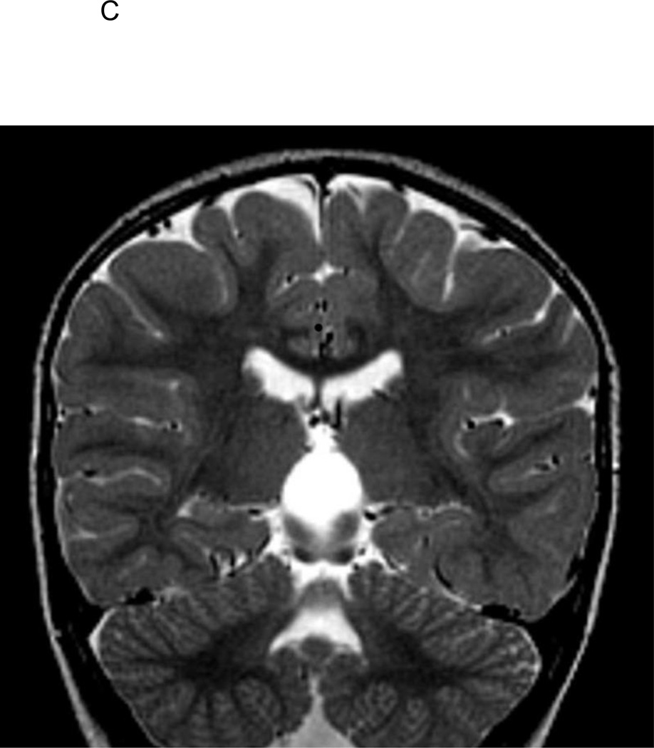
Figure 1.
A. T2-weighted coronal image of 2 1/2-year old FSE patient demonstrating medial positioning and globular shape of the left hippocampus.
B. T2-weighted coronal image of 2 1/2-year old FSE patient demonstrating medial positioning and globular shape of the left hippocampus, with associated vertically orientation of the left collateral sulcus.
C. T2-weighted coronal image of 2 1/2-year old FSE patient demonstrating medial positioning and globular shape of the left hippocampus, with associated inferior positioning of the posterior left crus of the fornix.


