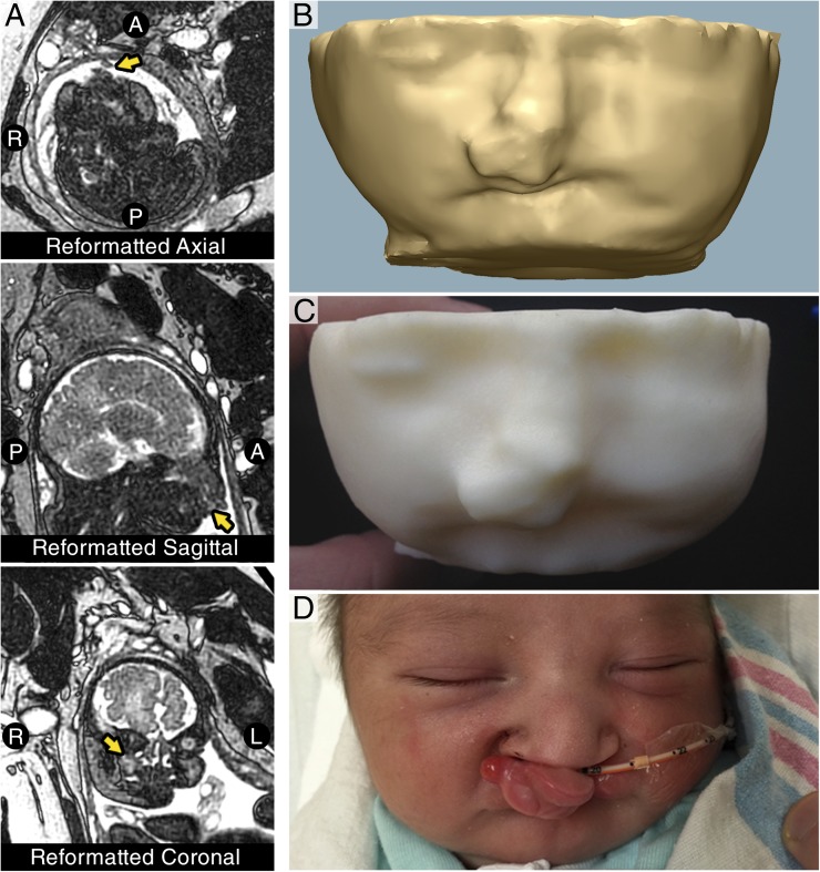
FIGURE 2.
Segmentation, modeling, and 3D printing of fetal MRI. (A) Representative single-shot T2 turbo spin echo-weighted MRI of the fetus reformatted in axial, sagittal, and coronal planes, which fail to clearly demonstrate the anatomy, making clinical decision-making difficult. (B) 3D model of the fetal facial soft tissues. (C) Final 3D-printed model of the fetal soft tissues demonstrating protuberant, isolated mass of the upper lip that does not involve the oral aperture. (D) Patient after successful delivery by cesarean delivery demonstrating bilateral cleft lip and palate with anteriorly displaced premaxilla.

