Figure 2.
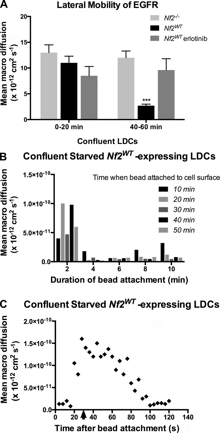
Rapid, local, signal-dependent immobilization of EGFR in confluent Nf2WT-expressing cells. (A) Lateral mobility of EGFR in confluent, serum-starved Nf2wt-expressing versus Nf2−/− LDCs at early (0–20 min) and late (40–60 min) time points after EGF-labeled beads were added. Also shown is the impact of 1-µM erlotinib treatment on EGFR mobility in Nf2WT-expressing cells. Data are represented as mean ± SEM. ***, P < 0.001 (one-way ANOVA with multiple comparisons). (B) Lateral mobility of EGFR in confluent, serum-starved Nf2WT-expressing LDCs measured at 2-min intervals after visually observing bead-to-cell attachment. Data are binned according to the time elapsed between exposing cells to EGF-labeled beads and observing bead-to-cell attachment. The graph shown is a representative time course of the mobility of a single bead over time. For each experiment, n > 3 beads. (C) Lateral mobility of single EGF-labeled beads on the surface of confluent, serum-starved Nf2WT-expressing cells at increasing time points after release from a laser optical trap (arrow). Data are representative of at least three experiments.

