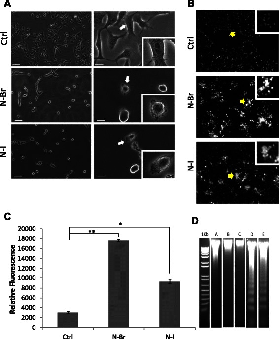
Fig. 2.

Benzofuroxan derivative effects on melanoma cells (a) Morphological effects. Arrows point to surface membrane alterations. Original magnifications, 100× left panels; 200× right panels; 400x inserts. b DNA condensation induced by N-Br and N-I. Yellow arrows indicate positive cells for Hoechst staining. 100×, and 400× inserts. c Quantification of Hoechst 33342 relative fluorescence in treated and control cells, shown in B; **p ≤ 0.001; *p ≤ 0.05 (d) DNA fragmentation showing a ladder pattern; A: Control; B: N-Br (16 μM); C: N-I (12 μM); D: N-Br (100 μM); E: N-I (100 μM)
