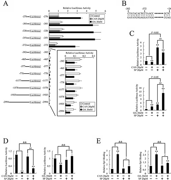
Figure 4. Identification of the CDK1 promoter regions which responded to PP2A inhibitors in PANC-1 cells.

A. Schematic diagram of the CDK1 promoter deletions created in the luciferase reporter construct, and the relative luciferase activities derived from the deletion constructs when transfected into PANC-1 cells. Cells were treated with PP2A inhibitors for 36 h after transfection and before the luciferase gene reporter assays were performed. The activity of the −276~+24 bp construct was given a value of 1, and the activities of the other transfections were normalized to this value. B. Sequence of the responsive element. C. JNK independent regulation of the activity of the −372~+24 bp construct; D. JNK dependent regulation of the activity of the −382~+24 bp construct, after treatment with PP2A inhibitors: after transfection, cells were treated with the JNK inhibitor for 3 h, and then treated with PP2A inhibitors for 36 h, followed by the luciferase gene reporter assays. E. JNK dependent regulation of binding between Sp1 and the responding element: cells were pretreated with SP600125 for 3 h, and then treated with PP2A inhibitors for 36 h, followed by the ChIP assays. *P < 0.05 and **P < 0.01 vs. respective control groups; #P < 0.05 and ##P < 0.01 vs. SP600125 group; &P < 0.05 and &&P < 0.01 induction between groups.
