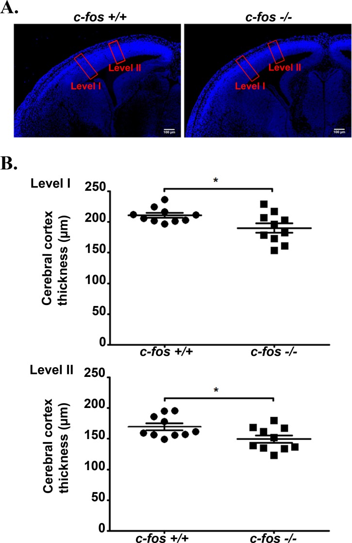
Figure 2. The neocortex of c-fos −/− embryos is thinner than that of their c-fos +/+ counterpart.
A. Fixed cerebral cortex of E14.5 c-fos +/+ and c-fos −/− embryos were stained with DAPI and examined under a confocal microscope. Boxes in red represent the areas used for the quantifications shown in B. B. The thickness of the neocortex was determined by measurements performed at two different right-left levels (I and II) for each genotype slice as marked in A. Four determinations were done in each sample; 10 samples of each genotype were examined. Results shown are the mean thickness of neocortex (μm) ± SEM. *p < 0.05 in c-fos −/− embryos with respect to the c-fos +/+ littermates using unpaired t test with Welch's correction.

