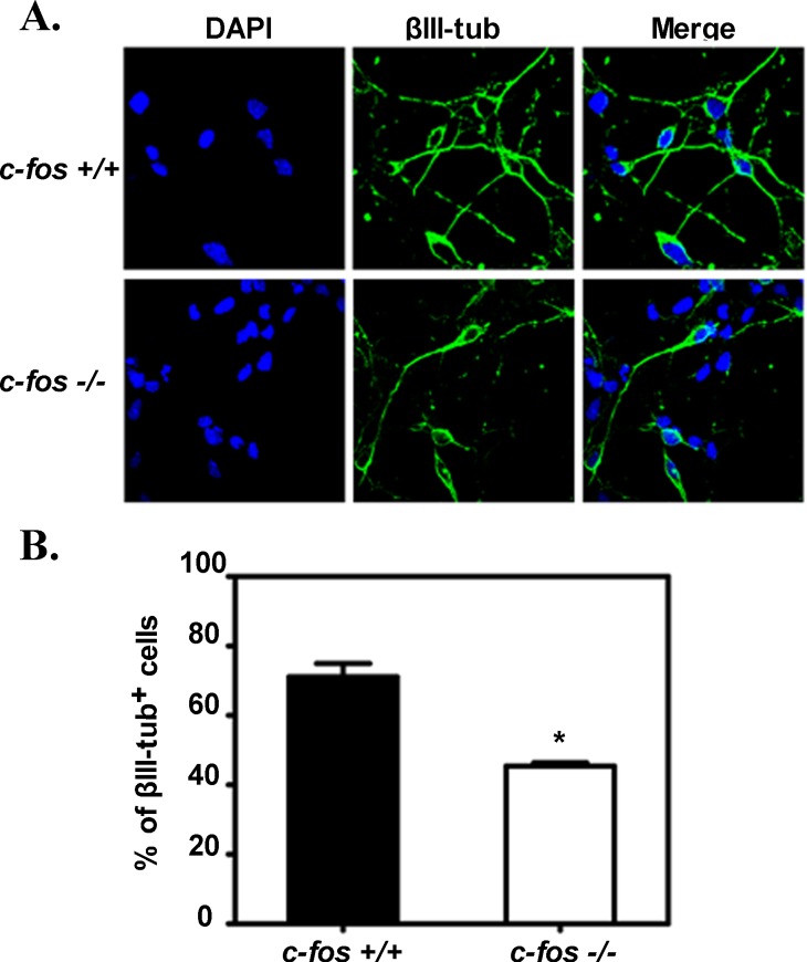
Figure 9. NSPCs from c-fos −/− cerebral cortex undergo less neuronal differentiation in the presence of NGF.
A. NSPCs isolated from cerebral cortex of E14.5 c-fos +/+ and c-fos −/− embryos induced to differentiate in vitro by culturing during five days in the presence of 40 ng/ml of NGF were analyzed by immunofluorescence for βIII-tubulin (green) and DAPI (blue) staining. B. Percentage of βIII-tubulin positive (+) cells shown in A. Results are the mean percentage values of cells showing positive staining for βIII-tubulin ± SEM calculated from the quantification of 10 fields from 3 different independent experiments. Number of cells with positive DAPI staining was considered as 100%. Statistical analysis: *p < 0.01 in NSPCs isolated from c-fos −/− embryos with respect to the c-fos +/+ condition as determined by unpaired t-test with Welch's correction.

