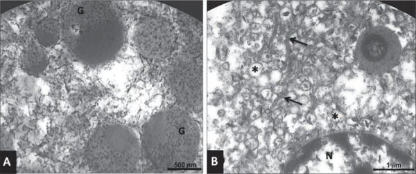
Figure 6. Transmission electron microscopy (TEM) of a human sublingual gland with advanced autolysis (male, 71 years old). A: Secretion granules of a serous cell (G) with partially preserved content and destruction of cytoplasmic organelles; B: Cisternae (arrow) and vesicles (*) of the rough endoplasmic reticulum and preserved nucleus (N) of a serous cell.