
Figure 6.
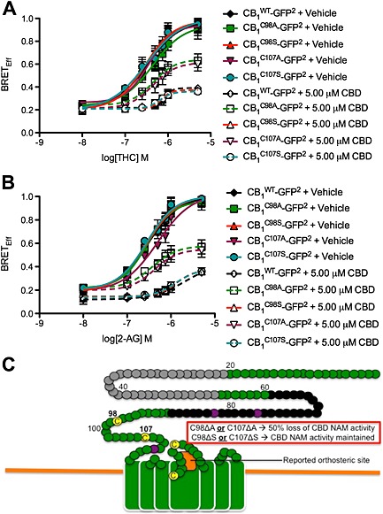
Cys98 and Cys107 coordinate the NAM activity of CBD at CB1 receptors. (A and B) STHdh Q7/Q7 cells were transfected with arrestin2‐Rluc‐ and CB1 C98A‐GFP2‐, and CB1 C98S‐GFP2‐, CB1 C107A‐GFP2‐ and CB1 C107S‐GFP2‐containing plasmids, and BRET2 was measured 30 min after treatment with THC (A) or 2‐AG (B) ± CBD. CRCs were fit using nonlinear regression with variable slope (four parameters) N = 4. (C) Diagram of the membrane‐proximal region of CB1 receptors summarizing data presented in this figure (adapted from Fay and Farrens, 2013). Our observations and previous studies suggest that Cys98 and Cys107 contribute to CB1 receptor allosterism, while the orthosteric site is near the second extracellular loop (orange box). In this diagram, green represents extracellular surface of CB1 receptors. Black circles represent residues unique to the N‐terminus of CB1A receptors Grey circles represent residues unique to the N‐terminus of CB1B receptors. Yellow circles represent Cys. Purple circles represent N‐glycosylated residues. Residues mutated in this study are marked in bold. Non‐bold numbers indicate amino acid number relative to N‐terminus.