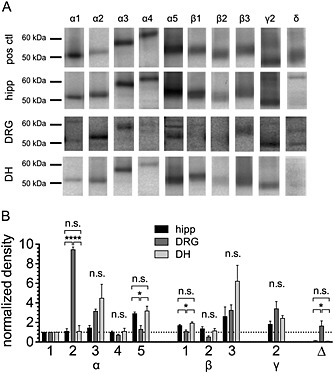
Figure 1.

Comparison of the expression pattern of GABAA receptor subunits in hippocampal, dorsal root ganglia and dorsal horn neurons. Hippocampus (hipp), dorsal horn (DH) and dorsal root ganglia (DRG) were collected from 10–14‐day‐old rats, and membrane proteins were prepared. The proteins were separated on 10% polyacrylamide minigels and transferred to membranes, which were then incubated in one of 10 different antibodies directed against various GABAA receptor subunits. As positive controls (pos ctl) showing reference positions and densities of bands, the same amount of membrane protein from whole mouse brain was used for the antibodies against the α1, α2, α3, α4, β1, β3 and γ2 subunits. For the antibody against α5, membrane proteins from mouse hippocampus were used, while mouse cerebellum was used as positive control for the antibodies directed against the β2 and δ subunits. (A) Shows the various bands obtained with the antibodies in membrane preparations of the three different tissues, as observed in one experiment. (B) The densities of all bands were normalized to that of the α1 band within the same tissue obtained in the same experiment; the results show the average values for three independent experiments. * and **** indicate significant differences at P < 0.05 and P < 0.0001 (ANOVA, followed by Holm–Sidak's multiple comparison post hoc test with pooled variance).
