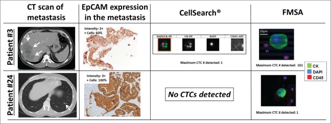
Figure 4.

Illustrated are CTCs isolated with CellSearch®, the FMSA, CT scans and EpCAM immunohistochemistry of resected metastases of 2 patients that underwent CRC liver (patients #3) and lung (patient #24) metastasectomy. CTC immunofluorescence staining images from the CellSearch® system and the FMSA, CT scans of liver and lung metastases, and EpCAM immunostaining (peroxidase method) and the respective expression intensity scores (0–3) and percentages of EpCAM+ tumor cells in resected and paraffin-embedded metastases are shown (from left to right column). EpCAM was consistently expressed in CRC liver and lung metastases at high levels, also in patients that had no CTCs detectable by EpCAM-based CellSearch® system (patient #24).
