
Figure 3.
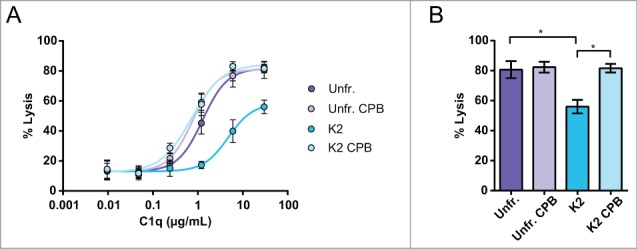
C-terminal lysine mediated inhibition of CDC results from less effective use of C1 when C1q was titrated in C1q depleted serum. (A, B; n = 3). The unfractionated (purple symbols and bars) and K2 isoform (blue symbols and bars) with and without CPB treatment were analyzed. Daudi cells were incubated at 37°C for 45 min and lysis was assessed by flow cytometry using a PtdIns exclusion assay. The level of CDC is expressed as percentage of total cells. The absence of CDC without added C1q indicates classical pathway activation. The data represents mean ± SEM and statistical significance was assessed for maximal lysis by one-way ANOVA followed by a Tukey post hoc test (*p < 0.05).