
Figure 4.
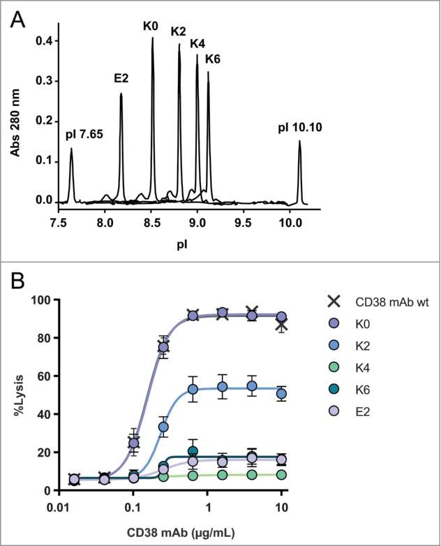
Overlay of capillary isoelectric focusing profiles of CD38 C-terminal mutants (A). The mutant abbreviations K0, K2, K4, K6 and E2 (Table S1) and pI markers 7.65 and 10.10 are indicated and detection occurred at 280 nm. Dose-response curves of CDC mediated cell lysis of the C-terminal mutants (n = 3) (B). NHS (20% vol/vol) was used as complement source and cells were incubated at 37°C for 45 min. Lysis was assessed by flow cytometry using a PI exclusion assay and the level of CDC is expressed as percentage of total cells. CD38 mAb wt produced in HEK-293F was used as positive control.