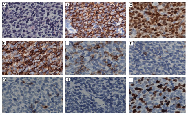
Figure 1.

Histopathology of testis. (A) Section from testis shows diffuse lymphoblasts with high nuclear/cytoplasm ratio, irregular shaped nuclei and indistinct nucleoli, obvious mitosis(H&E). (B, C, and D) positivity for CD20, TdT and CD43. E: part positive staining for CD34. (F, G, and H) negativity for CD3,CD7, MPO. I: About 30% of the neoplastic cells display nuclear Ki67 staining.
