
Figure 4.
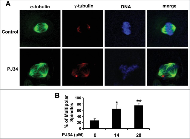
PJ34 induced multiple spindle formation in breast cancer BT549 cells. (A) BT549 cells were treated with PJ34 at 14 µM. After 24 h incubation, the cells were fixed, permeabilized and immunolabeled for α- and γ-tubulin for the detection of spindles and centrosomes (green and red fluorescent labeling, respectively). DNA was labeled with NucRed647 (blue). (B) The percentage of mitotic cells with multipolar spindles was calculated in BT549 cells treated with DMSO control or PJ34 at 14 and 28 µM. All the values are the average of triplicate determinations with the s.d. indicated by error bars. *P < 0.05, **P < 0.01 versus corresponding BT549 cells treated with DMSO control.