Figure 2.
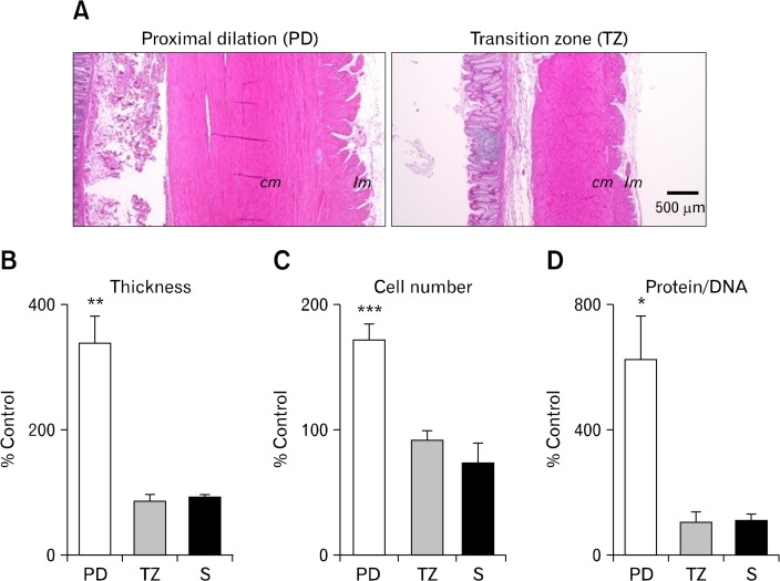
Morphometric characteristics of the smooth muscle layers. (A) H&E-stained cross-section demonstrating notable hypertrophy in the circular (cm) and longitudinal muscle (lm) layers in a proximal dilated (PD) colon (left panel) as compared with transition zone (TZ, right panel). (B) Cross-sectional muscle thickness (including both circular and longitudinal muscle layers) was significantly increased in the PD region in comparison with the control. (C, D) Smooth muscle hypertrophy was accompanied by an increase in the myocyte number as estimated by counting the nuclei (C), as well as an increase in myocyte size as measured by the protein-to-DNA content ratio (D) which indicates that the cell phenotype had been changed. S, non-dilated sigmoid colon. *P < 0.05, **P < 0.01, ***P < 0.001.

