Figure 2.
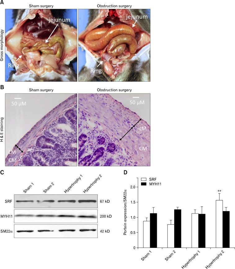
Gastrointestinal (GI) smooth muscle hypertrophy in mouse partial obstruction model. Partial obstruction related hypertrophy was surgically induced for ∼2 weeks by placing a small silicon ring on the distal ileum of mice. (A) Gross images of GI tract in sham (large ring) and obstruction (small ring) surgeries. (B) Representative H&E staining of jejunal cross sections from sham control and partially obstructed mice. Hypertrophied jejunum contained significantly thicker circular (CM) and longitudinal muscle (LM) layers compared to a sham control. (C) Western blot analysis of SRF, MYH11, and SM22α in hypertrophied and sham control jejunum. (D) Summary of Western blot analysis. Samples were run in triplicates for each animal (n = 2), and bars represent standard error of the mean. **P < 0.01.

