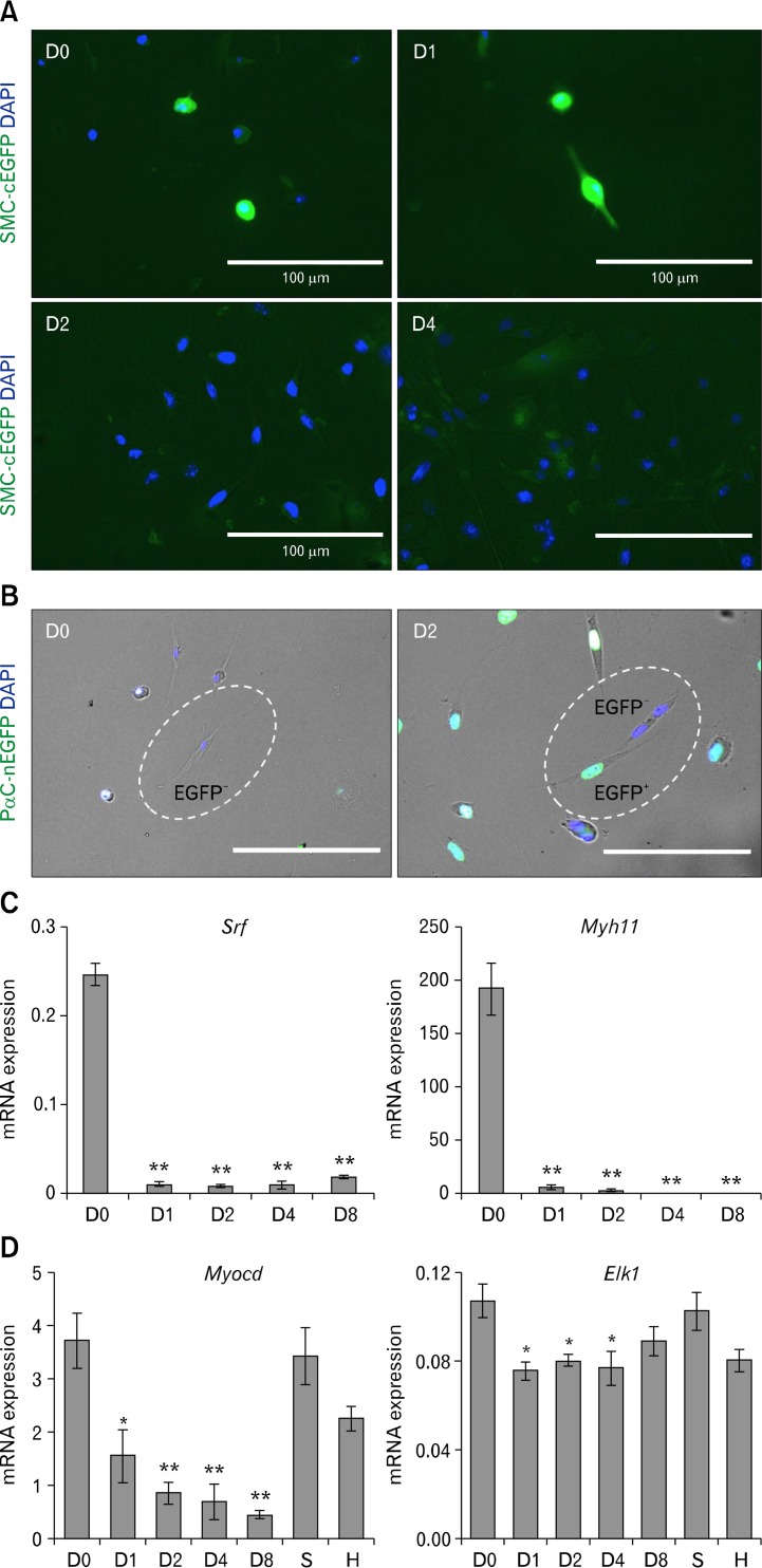
Figure 5.
Loss of Srf, Myocd, and Myh11 with gain of platelet-derived growth factor receptor alpha (PDGFRα) expression in dedifferentiated smooth muscle cells (SMCs) during cell culture. Jejunal smooth muscle was dissected from 2-week-old Myh11-Cre-EGFP or Pdgfra-EGFP mice, and the cells were dispersed and cultured for 8 days. At designated time points, cultured cells were examined by epifluorescence imaging and/or harvested for quantitative polymerase chain reaction (QPCR) analysis. (A) Epifluorescence imaging of SMCs from a Myh11-Cre-EGFP mouse showing loss of cEGFP (green) after day 2 in culture. DAPI (blue) marks nuclei in cells. (B) Epifluorescence imaging showing gain of nEGFP (green) expression in SMCs from a Pdgfra-EGFP mouse after 2 days in culture indicating activation of PDGFRα expression in SMCs. (C) QPCR analysis showing immediate and dramatic loss of Srf and Myh11 mRNA transcripts in primary SMCs after day 1 of culture. (D) QPCR analysis showing gradual loss of Myocd mRNA transcripts and sustained expression of Elk1 in primary SMCs after day 1 of growth in culture, in hypertrophic jejunum (H), and in sham controls (S). Expression of each gene was normalized by housekeeping gene, Gapdh (n = 3). *P < 0.05, **P < 0.01.

