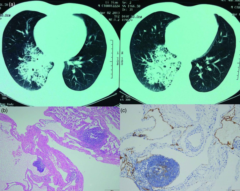
Figure 1:
(a) High-resolution CT scan demonstrating high-density opacity and interlobular septal thickening especially in the right lower lobe of the lung. (b) Microscopic examination showing diffuse pulmonary lymphangiomatosis located in the right middle and lower lobe. Hematoxylin and eosin-stained section of the lesion showing that proliferative lymphatic channels were diffusely infiltrative along the intralobular septum without cytological atypia (×200). (c) Immunohistochemical staining with D2-40 revealing proliferative lymphatic channels (×200).

