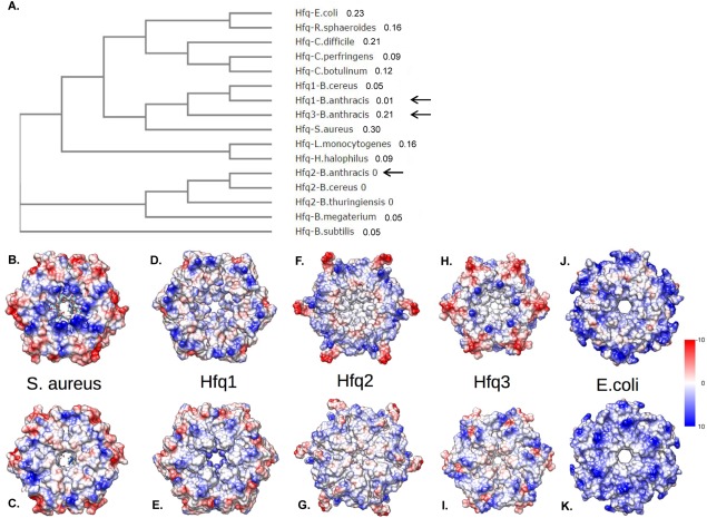
Figure 2.
Bacillus anthracis Hfq phylogeny and modeling. (A) Unrooted cladogram depicting relationship of Hfq1-3 protein sequences (black arrows) with those of other bacterial species. Numbers designate branch lengths. (B–K) Models of Hfq hexamers. Upper row depicts proximal face and lower row depicts distal face for the crystal structure of S. aureus Hfq (B,C); models of B. anthracis Hfq1, 2, and 3 (D–I); or the crystal structure of E. coli Hfq (J,K). Surfaces are colored by electrostatic surface potential, from blue (highly positive) to red (highly negative).

