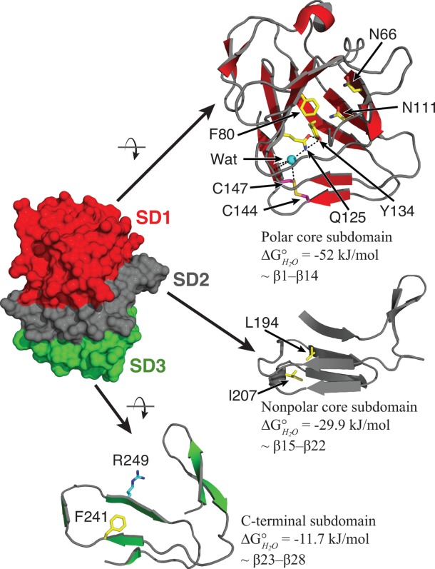
Figure 4.

Subdomain divisions and residues mutated within HpmA265. Protein subdomains are as discussed in the text and are colored as polar core subdomain (red), nonpolar core subdomain (gray), C-terminal subdomain (green). The internal amino acid residues mutated in this study are shown as sticks colored by atom type (yellow – carbon; blue – nitrogen; red – oxygen; magenta – sulfur). Carbon atoms on R249, the site of the trypsin cleavage, are colored cyan. Black dashed lines indicate probable hydrogen bonds involving Q125, Y134, and an internal water (Wat, cyan sphere).
