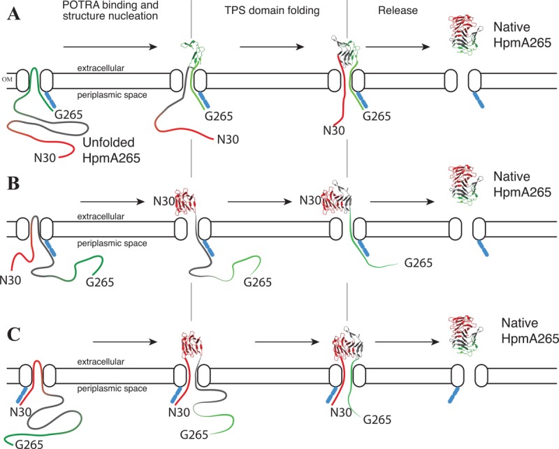
Figure 6.

TPS domain driven secretion schemes for HpmA265. Unfolded HpmA265 is shown as a ribbon that is colored by subdomain: polar core (red), nonpolar core (gray), C-terminal (green). The N- and C-terminus are labeled. The outer membrane is shown with an embedded TpsB component. Secretion and folding proceed from left to right in each panel. The POTRA domains are found on the periplasmic side and are represented by blue ovals. Three possible transport schemes are shown. (A) POTRA domains bind C-proximally on HpmA265 and remain bound until translocation is (nearly) complete. (B) POTRA domains binds initially near the middle of HpmA265. As secretion progresses, the POTRA domains are bound in progressively more C-proximal locations. (C) POTRA domains bind N-proximally on HpmA265 and remain bound until translocation is (nearly) complete.
