Figure 2.
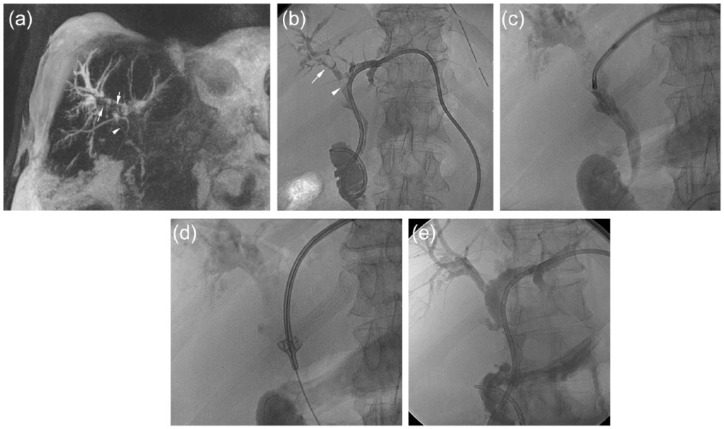
A 64 year-old man with positive history for primitive sclerosant cholangitis, portal thrombosis and cholecystectomy presented with ascites, recurrent cholangitis and intrahepatic lithiasis. (a) Magnetic resonance (MR) T2w IR in coronal view shows bilateral stenosis of the bile ducts with presence of biliary stones in the right bile duct (arrows) and in the CBD (arrowhead) (Tsunoda class IV). Moreover abundant ascites is present around the liver. (b) A left transhepatic approach through the bile duct of the III segment was performed and a biliary drainage was placed. Cholangiography confirmed the MR findings: intrahepatic lithiasis (arrow) and lithiasis in the CBD (arrowhead). (c,d) Cholangioscopy was performed using a 11 Fr introducer with the small cholangioscope (size 2.7 mm) and also using a basket-type catheter. (e) After 3 days, cholangiography shows the optimal result without any sign of residual biliary stones.

