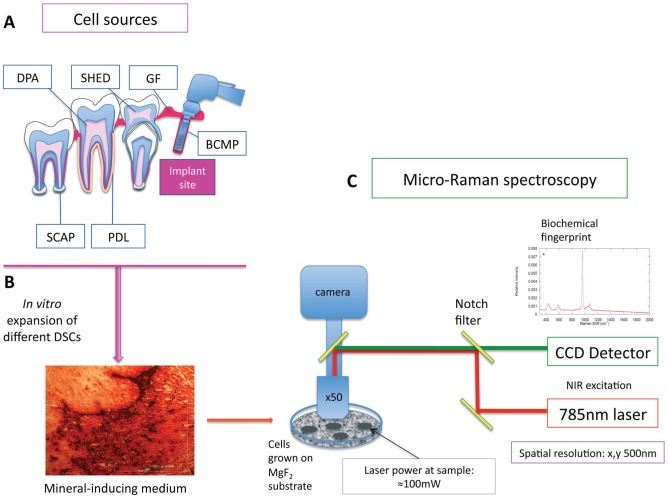
Figure 1.
Schematic representation of pathway of analysis of cell populations. Different mesenchymal cells were isolated from different dental and supporting tissues (A). After their in vitro expansion, the cells were grown in mineral-inducing media for 28 d. All cells formed minerals in vitro that were confirmed with alizarin red staining, marking the calcium deposition (B). Cells were grown on MgF2 coverslips (to facilitate Raman spectral analysis) and analyzed by micro-Raman spectrometer outfitted with a Leica microscope and 785-nm in-line diode laser (C). BCMP, bone chip mass population; CCD, charge-coupled device; DPA, dental pulp adult; GF, gingival fibroblast; NIR, near infrared; PDL, periodontal ligament; SCAP, stem cells from apical papilla; SHED, stem cells from human-exfoliated deciduous teeth.

