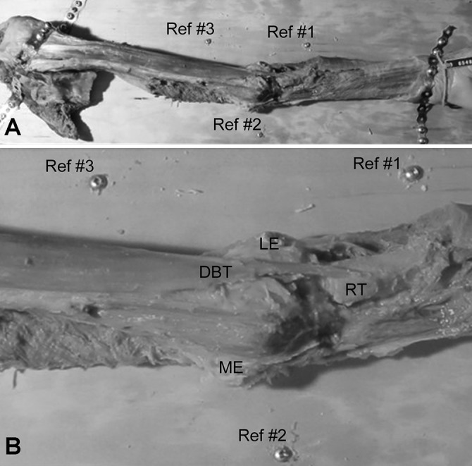
Figure 1.

(A) Photograph illustrating the specimen setup for a left elbow. The limb was dissected and secured to a wooden board using metal plates and screws (scapula to the left, hand to the right). The elbow was positioned in full extension with the forearm maximally supinated. Three reference screws (Ref #1-#3) were positioned about the elbow. (B) An enlarged photograph from above the distal biceps brachii muscle and tendon. DBT, distal biceps tendon; LE, lateral epicondyle; ME, medial epicondyle; RT, radial tuberosity.
