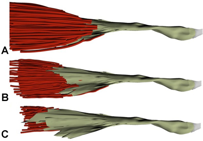
Figure 4.

A 3-dimensional representation of the distal muscle tendon unit of a single left elbow specimen chosen at 3 distinct times during the digitization process. (A) Prior to any dissection of the muscle layers, (B) after about half of the muscle fibers have been dissected off the tendon, and (C) after the majority of the muscle fibers have been dissected off the tendon, revealing the full length of the internal tendon structure.
