Figure 1.
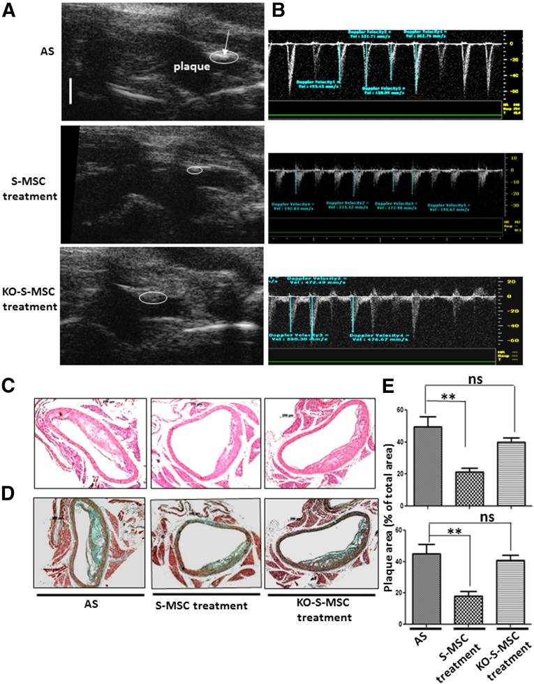
Effects of S-MSC treatment on established atherosclerotic plaque progression in brachiocephalic arteries of apolipoprotein E-knockout (apoE−/−) mice. We used a mouse model for AS in apoE−/− mice and constructed three groups of mice: an AS (control group) comprising apoE−/− mice fed a high-fat diet for 24 weeks, and another two groups of apoE−/− mice that were fed a high-fat diet for 24 weeks and received 0.5 million wild-type S-MSCs or KO-S-MSCs weekly injections for the final 5 weeks. Ultrasound biomicroscopy (was performed after 24 weeks of treatment. Representative ultrasound biomicroscope (UBM) images showed the brachiocephalic arteries of the three groups of mice (AS, S-MSC treatment, KO-S-MSC treatment). (A): S-MSC treatment reduced the plaque area in brachiocephalic arteries of apoE−/− mice. White bar for UBM images = 1 mm. (B): Images of pulse-wave Doppler measurements of maximal flow velocities in the corresponding groups. (C, D): H&E (C) and Masson trichrome staining (D) show histopathologic changes of the mouse aortic arch. (E): Quantitative analysis of lesion size in the three groups. All results shown are representative of the arithmetic mean ± SEM of eight independent experiments. Scale bar for histological images = 200 µm. ∗∗, p < .01 versus AS. Abbreviations: AS, atherosclerosis; KO-S-MSCs, nuclear factor-κB−/− skin-derived mesenchymal stem cell treatment group; ns, nonsignificant; S-MSC, skin-derived mesenchymal stem cell.

