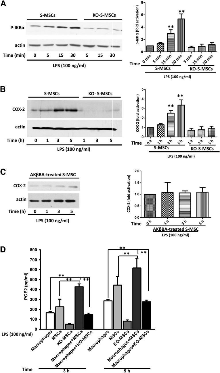
Figure 4.
Examination of molecular alterations underlying the effect of S-MSCs on macrophages. (A, B): Wild-type S-MSCs or KO-S-MSCs (1 × 10⁶ per assay) were stimulated with LPS (100 ng/ml) for the indicated time. Whole-cell lysates were separated and analyzed by Western immunoblots with antibodies phosphor-IκBα (A) and COX-2 (B). (C): Effects of NF-κB on the LPS-induced COX-2 expression. S-MSCs pretreated with 10 µmol/l AKβBA for 30 minutes were stimulated with LPS (100 ng/ml) for the indicated time on the immunoblot with staining of actin-loading controls. Densitometric scanning of three Western blots is quantified in the bar graphs. The results shown are representative of at least three independent experiments. (D): S-MSCs isolated from wild-type or NF-κB−/− mice were grown to confluence and cultured or cocultured with macrophages for an additional period of 3 or 5 hours with LPS (100 ng/ml) stimulation. Supernatants were collected, and the levels of PGE2 were determined by enzyme-linked immunosorbent assay (n = 4). Error bars represent mean ± SEM. ∗∗, p < .01. Abbreviations: AKβBA, acetyl-11-keto-β-boswellic acid; COX-2, cyclooxygenase-2; KO-S-MSCs, nuclear factor-κB−/− skin-derived mesenchymal stem cell treatment group; LPS, lipopolysaccharide; PGE2, prostaglandin E2; S-MSC, skin-derived mesenchymal stem cell.

