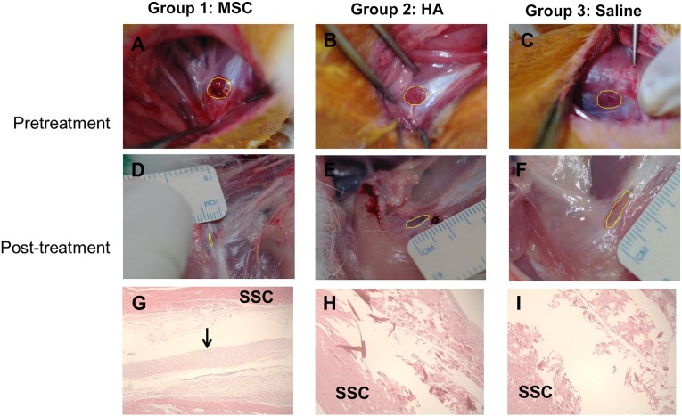
Figure 4.
Gross morphological (A–F) and histological (G–I) findings of the subscapularis tendons in groups 1, 2, and 3. The polygon in each of the first six images depicts the area of the full-thickness subscapularis tendon tear. (A–C): Pretreatment images. (D–F): Posttreatment images. (G): Parallel arrangement of hypercellular fibroblastic bundles (arrow) was noted in group 1. (H, I): Histological findings in groups 2 and 3 showed absence of fiber bundles. Group 1 received a 0.1-ml injection of MSCs; group 2, 0.1 ml of HA; group 3, 0.1 ml of saline. Hematoxylin-and-eosin stain, ×40. Abbreviations: MSC, human umbilical cord blood-derived mesenchymal stem cell; HA, hyaluronic acid; SSC, subscapularis muscle.

