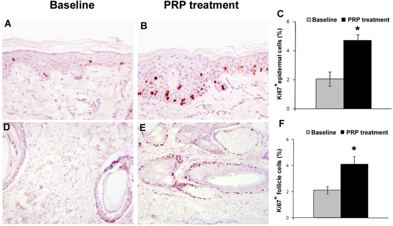
Figure 3.
Proliferation of epidermis basal cells and hair follicular bulge cells is increased after PRP treatment. (A, B): Representative photomicrographs of Ki67+ proliferating cells by immunohistochemistry of hair skin epidermis at baseline (A) and after PRP treatment (B) (hematoxylin and eosin stain; original magnification: ×200). (C): Morphometric analysis of Ki67+ cells of hair skin epidermis at baseline and after PRP treatment. (D, E): Representative photomicrographs of Ki67+ proliferating cells by immunohistochemistry of hair follicles at baseline (D) and after PRP treatment (E) (hematoxylin and eosin stain; original magnification: ×100). (F): Morphometric analysis of the percentage of Ki67+ nuclei in hair follicles at baseline and after PRP treatment. ∗, p < .05. Abbreviation: PRP, platelet-rich plasma.

