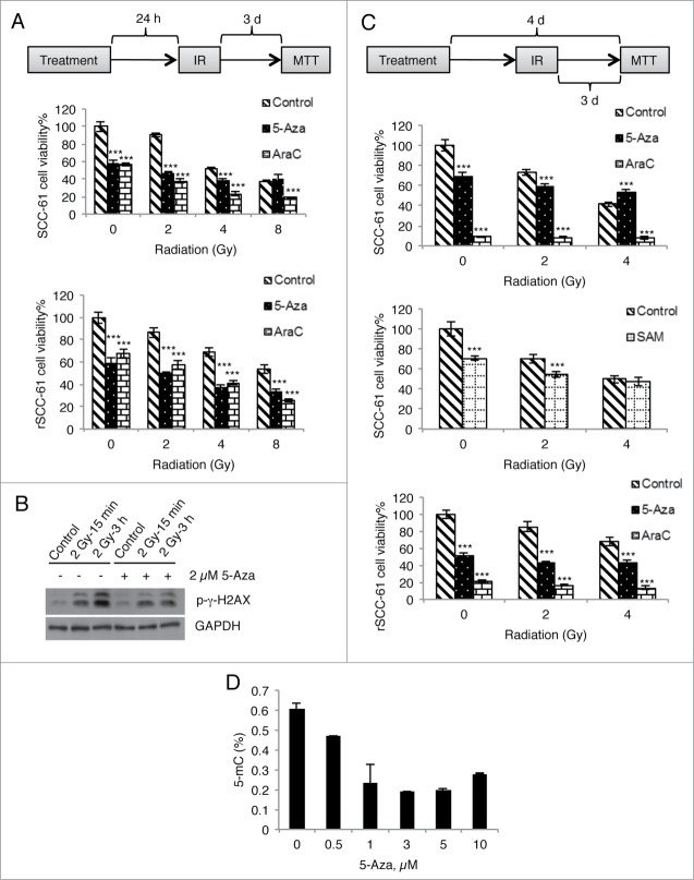
Figure 8 (See previous page).
Treatment with 5-Aza does not enhance radiation-induced DNA damage and cell death. (A) SCC-61 and rSCC-61 cells were incubated with 2 µM of 5-Aza or AraC overnight and then irradiated with a single dose of 2, 4, or 8 Gy. Cell viability was determined at 72 h post-irradiation using an MTT assay. (B) rSCC-61 cells were incubated with 2 µM 5-Aza for 4 days and then irradiated with a single dose of 2 Gy. Cell lysates were collected at 15 min and 3 h post-irradiation and subjected to Western blot analysis using antibodies against p-γ-H2AX and GAPDH. (C) SCC-61 and rSCC-61 cells were incubated with 2 µM 5-Aza, 2 µM AraC, or 100 µM SAM for 4 days (refreshing the medium every other day), and treated with a single dose of 2 or 4 Gy irradiation starting on the second day. Cell viability was determined at 72 h post-irradiation using an MTT assay. In both (A) and (C), asterisks indicate statistically significant changes in cell viability for 5-Aza- and AraC-treated cells at each radiation treatment condition relative to the DMSO control [α = 0.05, P-values of 0.01–0.05 (*), 0.001–0.01 (**), or <0.001 (***)]. (D) rSCC-61 cells were incubated with 0–10 µM 5-Aza for 4 days and 5-methylcytosine levels were measured using MethylFlash Methylated DNA Quantification Kit. Increasing concentrations of 5-Aza decreased global DNA methylation. Data are from 2 independent experiments. Asterisks indicate statistically significant changes [α = 0.05, P-values of 0.01–0.05 (*), 0.001–0.01 (**), or <0.001 (***)].

