Figure 4.

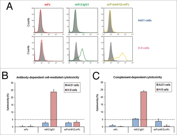
Summary of ADCC and CDC activity in H9 and A431 cells. (A) Mesothelin-negative A431 cells (left panel) and mesothelin-positive H9 cells (right panel) were incubated with 0.3 μM negative scFv (gray), mFc (red), m912 IgG1 (green) and scFv(m912)-mFc (gold). The cells were washed and further incubated with FITC-conjugated goat F(ab’)2 anti-human IgG, then washed and analyzed by flow cytometry. (B) IgG1 m912 induced ADCC in mesothelin-positive H9 cells but not in mesothelin-negative A431 cells. The scFv(m912)-mFc fusion protein did not induce detectable cytotoxicity. (C) IgG1 m912 induced CDC in mesothelin-positive H9 cells but not in mesothelin-negative A431 cells. The scFv(m912)-mFc fusion protein did not induce detectable cytotoxicity. The detection limit of the assays is indicated (dotted line) and represents 5% of lysis. Error bars show s.d. from an average of three separate experiments.
