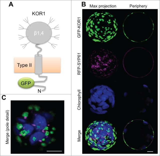
Figure 1.

GFP-KOR1 patterns at the surface of KOR1-deficient Arabidopsis rsw2–1 protoplasts upon co-expression of TGN marker RFP-SYP61. (A) Membrane topology of the GFP-KOR1 reporter protein. (B) Maximum (Max) projection of about 30 sections (left) and a central section (right) are shown for single and merged channels. (C) Pole detail of the same cell (Max projection of merged signals). Fluorescent signals were recorded ca. 48 h post-transfection. Green, GFP-KOR1; magenta, TGN marker RFP-SYP61; blue, chlorophyll. EG, endoglucanase domain; N, N-terminus. Bars represent 5 5 μm.
