
Figure 5.
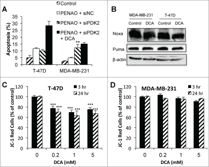
DCA can enhance apoptosis via off-target mechanisms. (A) Apoptosis in T-47D and MDA-MB-231 cells after 48 hr 2 μM PENAO treatment +/− 5 mM DCA in control (siNC) and siPDK2 cells (PDK protein expression 48 hr post-transfection with siPDK2 are shown in Fig. 2). (B) Noxa and Puma expression after 24 hr 5 mM DCA treatment. (C and D) Mitochondrial membrane potential (JC-1 red indicates presence of MMP) in (C) T-47D cells and (D) MDA-MB-231 cells after 3 and 24 hr treatment with 0.2–5 mM DCA. **P < 0.01 vs PENAO + siNC; *** P < 0.001 vs control.