Figure 1.

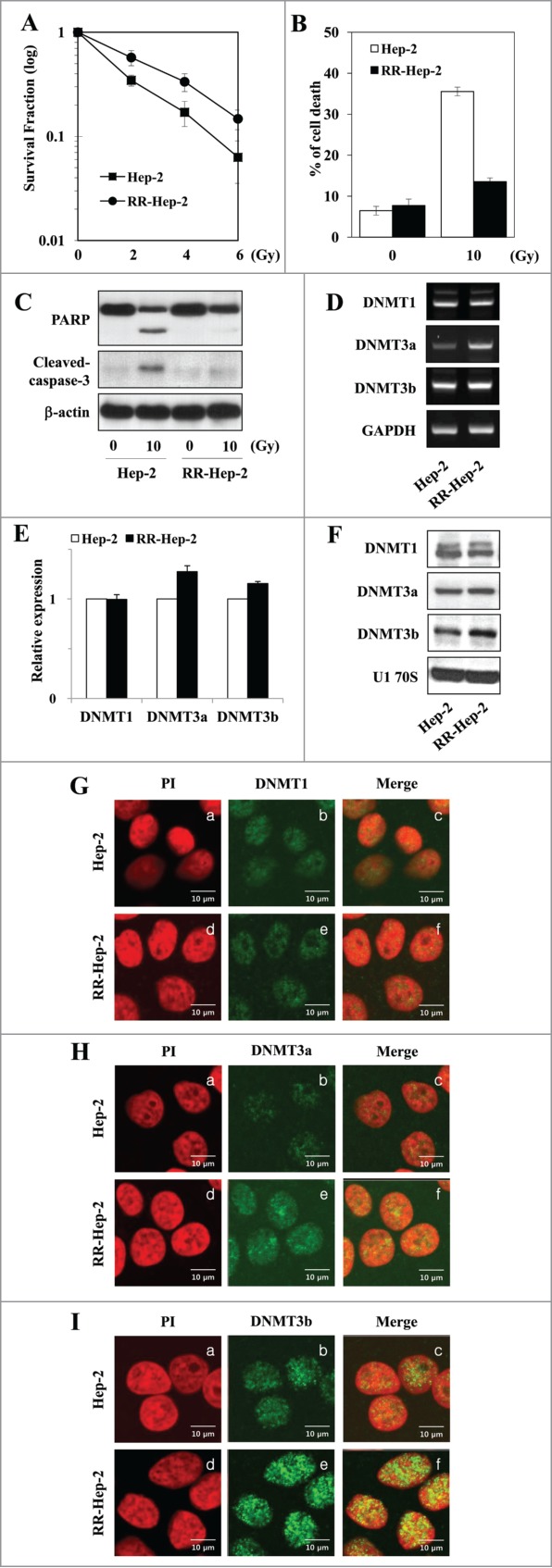
Analysis of survival and DNMT expression in our radioresistant Hep-2 human laryngeal cancer cell line (RR-Hep-2). (A) Clonogenic survival fractions of parental Hep-2 and RR-Hep-2 cells were determined following exposure to the indicated doses of radiation. Parental Hep-2 and RR-Hep-2 cells were treated with or without 10 Gy radiation and then cultured for 24 h. (B) Cell viability was determined with the FACScan flow cytometer and data are presented as the percentage of PtdIns-positive cells. (C) The protein levels of cleaved PARP and cleaved caspase-3 were determined by Western blotting; β-actin was employed as the loading control. (D) The transcript levels of DNMT1, DNMT3a and DNMT3b were determined by conventional RT-PCR and (E) quantified by real time PCR. GAPDH was used as the loading or internal control. (F) The protein levels of DNMT1, DNMT3a and DNMT3b were determined by Western blotting; U1 70S was employed as the loading control. (G-I) The localizations of (G) DNMT1, (H) DNMT3a and (I) DNMT3b were examined by immunofluorescence staining using an FITC-conjugated secondary antibody and propidium iodide staining (PI, for nuclei) followed by confocal microscopy. Panels: (a and d) PtdIns (red) staining; (b and e) localization of DNMTs (green); and (c and f) merged images of DNMTs and PI. Data are presented as results of a typical experiment from 4 independent experiments. The error bars represent standard deviations.
