Figure 5.

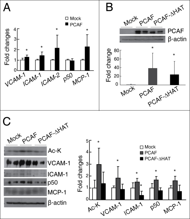
PCAF overexpression induces the expression of inflammatory molecules in cultured renal tubular cells. (A) qPCR analysis for VCAM-1, ICAM-1, ICAM-2, NF-κB p50, and MCP-1 in HK-2 cells transfected with either a pCI plasmid (Mock) or a pCI-Flag-PCAF plasmid (PCAF). (B) Representative western blots for PCAF and β-actin in the HK-2 cells transfected with a pCI plasmid (Mock) or a pCI-Flag-PCAF plasmid (PCAF) or a pCI-Flag-PCAF-ΔHAT plasmid (PCAF-ΔHAT) shown on the up panel, with their quantitative densitometric results shown on the bottom panel. (C) Representative western blots for Ac-K, VCAM-1, ICAM-1, NF-κB p50, MCP-1, and β-actin in HK-2 cells transfected with Mock, PCAF, or PCAF-ΔHAT shown on the left panel, with their quantitative densitometric results shown on the right panel. (Each experiment was performed in duplicates for 3 times with a representative result shown. *P < 0.05 compared with the Mock group).
