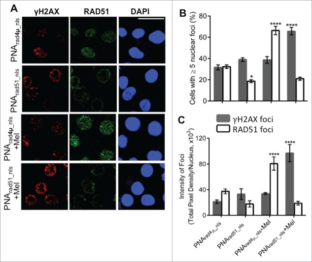
Figure 3.
PNA targeting RAD51 reduces de novo nuclear RAD51 foci and those induced by melphalan, and potentiates melphalan-induced DNA damage. (A, B) H929 cells were treated as indicated in “Materials and Methods.” Cytospins were made and stained for RAD51 and γH2AX. (A) Immunofluorescence pictures of cells after staining for RAD51, γH2AX and DAPI. (B) Quantitation of cells with ≥5 discrete nuclear foci of γH2AX or RAD51. At least 100 nuclei were analyzed for each treatment and nuclei with ≥5 foci were scored as positive. (C) Mean fluorescence (integrated pixel intensity per nucleus) ±SEM, of γH2AX and RAD51 foci after the indicated drug exposures. Data are shown as mean ± SEM of 3 experiments. * P < 0.05 ****P< 0.0001, for comparisons to controls. Prad4μ_nls, Prad51_nls and Mel indicate PNArad4μ_nls, PNArad51_nls and Melphalan, respectively. The scale bar (—) is 20 μm.

