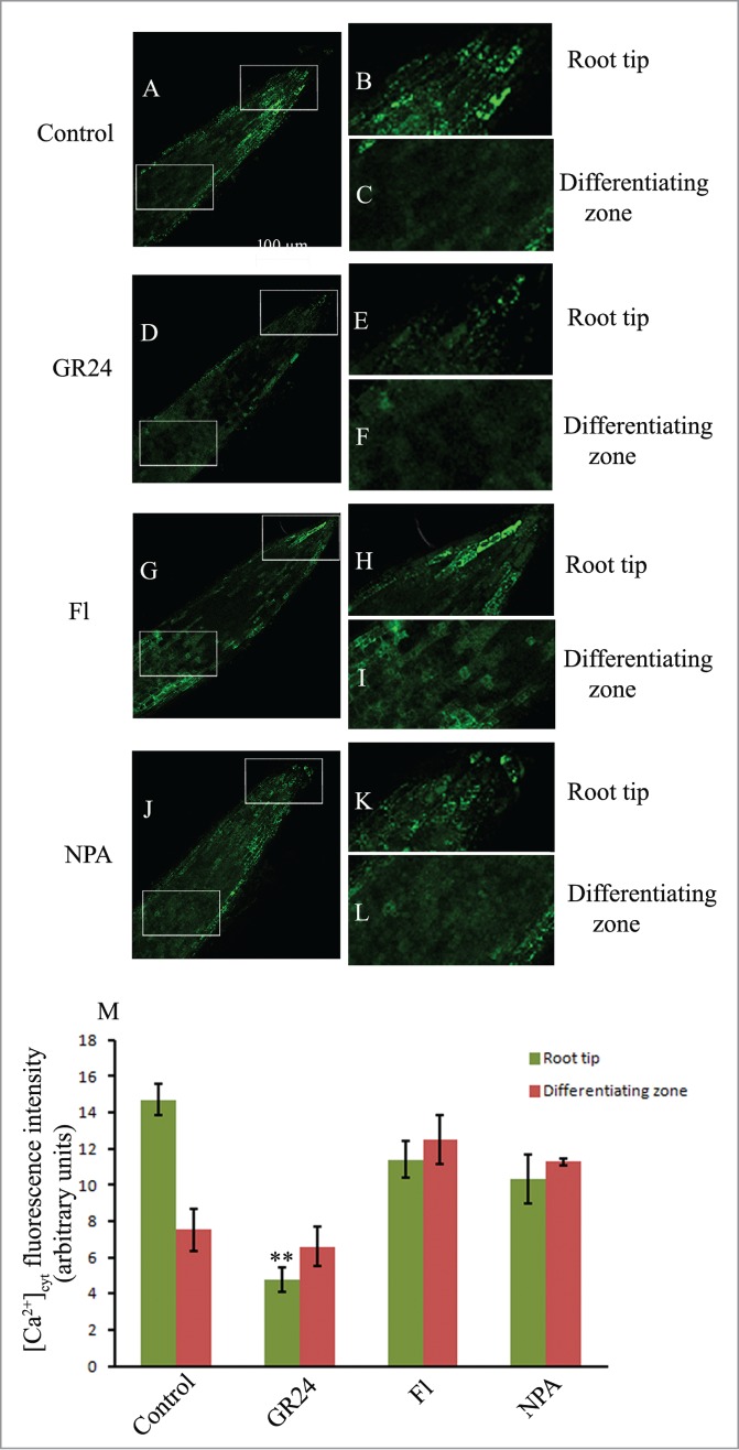
Figure 3.

CLSM analysis of cytosolic calcium ion [Ca2+]cyt distribution using Oregon green BAPTA-1 in the lateral roots of 6d old seedling grown in the distilled water (control; A–C) and other pharmacological agents (GR24, Fl, NPA; D–L). Magnification 20X; scale bar = 100 µm. Histogram shows relative fluorescence intensity of calcium ion distribution in the respective samples (M). Each datum indicates mean ± SE from at least 3 replicates, showing changes to be significant at different levels (*P < .05, **P < .01, ***P < .001) from the control, analyzed by one-way ANOVA.
