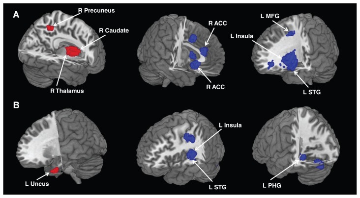
Fig. 2.
Areas of increased (red) and decreased (blue) resting-state brain activity in medication-free patients with major depressive disorder compared with healthy controls in the meta-analyses of (A) regional cerebral blood flow studies and (B) regional homogeneity studies. ACC = anterior cingulate cortex; L = left; MFG = middle frontal gyrus; PHG = parahippocampal gyrus; R = right; STG = superior temporal gyrus.

