
Figure 1.
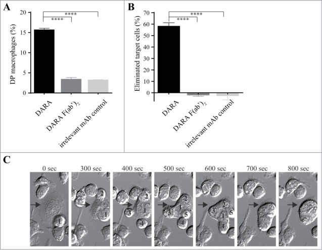
Flow cytometry and live cell imaging reveals macrophage-mediated phagocytosis of CD38+ tumor cells in the presence of DARA. Co-cultures of mouse mφ and Daudi cells in the presence of 6.7 nM DARA or F(ab’)2 fragments thereof, E:T ratio of 1:1 (A, B) or 3:1 (C). (A) Double positive (DP) mφ were characterized as F4/80+calcein+CD19– and the percentage DP macrophages was calculated as described in Materials and Methods. (B) The percentage eliminated target cells was calculated from the number of remaining F4/80- cells as described in Materials & Methods. Each bar shows mean ± SEM, results from a representative experiment are shown (n = 3). (C) Time-lapse imaging microscopy, bright field images of a mouse mφ (arrow) that sequentially engulfed 5 individual Daudi cells (numbers) over a period of 800 s. The images are representative for observations in multiple independent phagocytosis experiments (n = 3) (****p < 0.0001 Bonferroni's multiple comparison test).