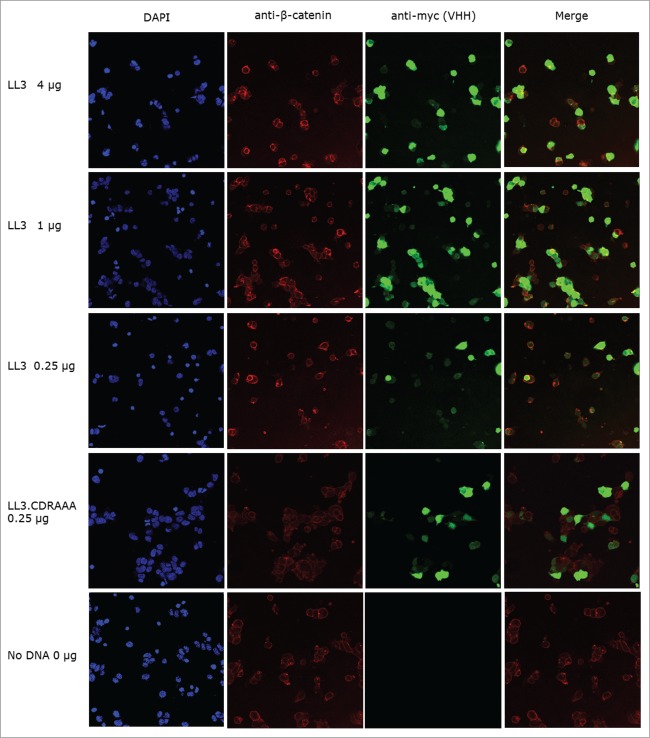
Figure 7.
Confocal microscopy of anti-β-catenin and intracellular antibody stained HEK293 bioassay cells. Fixed HEK293 bioassay cells were stained for intracellular antibody and β-catenin expression; the nuclear stain DAPI was also included. Anti-β-catenin and anti-intracellular antibody images were merged, and yellow donates co-localization. Pictures were taken using Leica TCS SP5 microscope, with 40× magnification.

