
Figure 1.
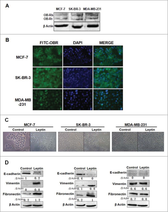
Leptin induces epithelial-mesenchymal transition of breast cancer cells. (A) Proteins were extracted from MCF-7, SK-BR-3 and MD-MB-231 cells. Western blotting analysis using specific antibodies demonstrated that Ob-Rb (125 kDa) and Ob-Rt (100 kDa) were expressed in the breast cancer cells. (B) Immunofluorescence assay with specific antibodies demonstrated leptin receptors were presented on the membrane of breast cancer cells. Original magnification: × 400 (C) The pictures of microscope showed EMT-related morphological changes. The presence of cell-cell junction dissolution, loss of apical-basolateral cell polarity emerged in MCF-7 and SK-BR-3 cells treated with leptin after 16 h serum starvation, while variations of morphological features were not observed in MD-MB-231 cells. Original magnification: ×200 (D) Westren blot analysisdemostrated that leptin downregulated E-cadherin, while upregulated Vimentin and Fibronectin of MCF-7 and SK-BR-3 cells. The EMT-related proteins in MD-MB-231 cells were not investigated.