
Figure 2.
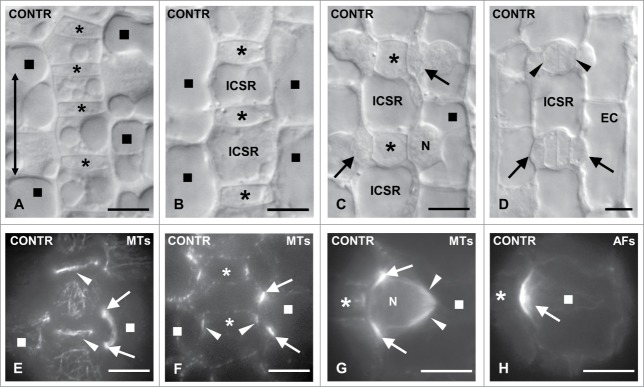
(A–D) Areas of Z. mays stomatal rows, as observed with DIC optics, displaying young GMCs (A), GMCs in an intermediate developmental stage (B), advanced GMCs (C) and young stomatal complexes (D). The double arrow in (A) shows the longitudinal leaf axis. The asterisks mark the GMCs and the squares the SMCs. In the SMC shown in (C) the nucleus (N) has occupied its polar position. The arrows point to the daughter cell walls of the asymmetrical divisions that create the subsidiary cells. The arrows in (D) show subsidiary cells and the arrowheads young guard cells. EC: epidermal cell; ICSR: intervening cell of the stomatal row. (E and F) GMCs (asterisks) and SMCs (squares) after tubulin immunolabeling in external (E) and median (F) optical planes. The arrows point to preprophase MT- band profiles of a SMC and the arrowheads to those of an interphase MT-ring in GMCs. (G) Tubulin immunolabeling in a prophase SMC (square). The arrows indicate the preprophase MT- band, the arrowheads the half prophase MT-spindle, while the asterisk marks the inducing GMC. N: nucleus. (H) AF-patch (arrow) in SMC (square), which bulges toward the inducing GMC (asterisk). Scale bars: 10 μm.