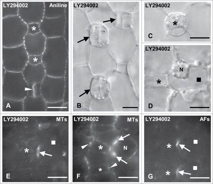
Figure 9.

Protodermal areas of seedlings incubated with LY294002. (A and B) Optical sections of stomatal rows after aniline blue staining (A) and under DIC optics (B). The asterisks mark advanced GMCs, the arrows young stomatal complexes and the arrowhead a newly formed daughter cell wall of the symmetrical GMC division. Note the absence of subsidiary cells in all cases. (C and D) Persistent GMC (asterisk in C) and treated SMC (square in D) as they appear under DIC optics. The SMC nucleus (N) has not occupied a polar position near the neighboring GMC (asterisk; cf. Fig. 2C). (E and F) Treated SMC (square) after tubulin immunolabeling in external (E) and median (F) optical planes. The arrows point to the SMC preprophase MT-band and the arrowheads show the GMC interphase MT-ring. N: nucleus. (G) Treated SMCs (squares) after AF staining. The arrows show the AF-patch in each SMC and the asterisks the neighboring GMC. Note the local "protrusion" of the SMCs toward the neighboring GMCs. Treatments: LY294002 50 μM, (A, D–G): 48; (B and C): 72 h. Scale bars: 10 μm.
