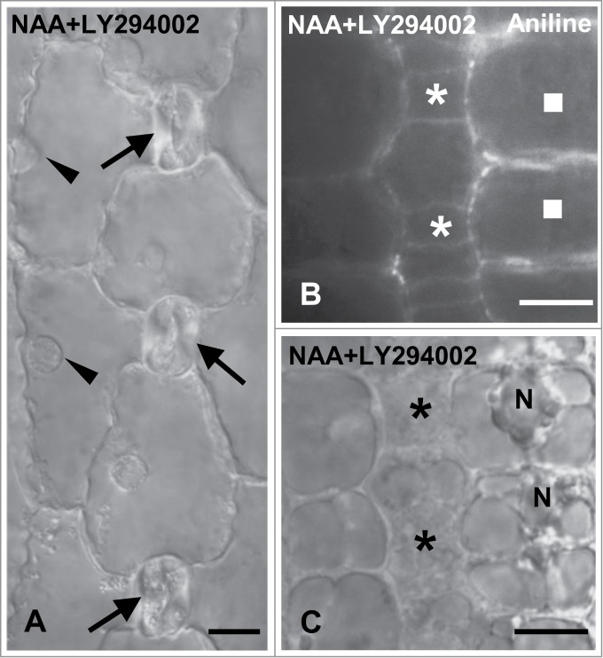
Figure 10.

Protodermal areas of seedlings incubated with NAA plus LY294002. (A) DIC optical view of a stomatal row. The young stomata (arrows) lack subsidiary cells. The arrowheads indicate the nucleus of each SMC that is located far from the respective stoma. (B and C) Treated SMCs (squares) after aniline blue staining (B) and in DIC optics (C). The nucleus (N) resides far from the adjacent GMCs (asterisks). Treatments: NAA 100 μM + LY294002 50 μM, 48 h. Scale bars: 10 μm.
