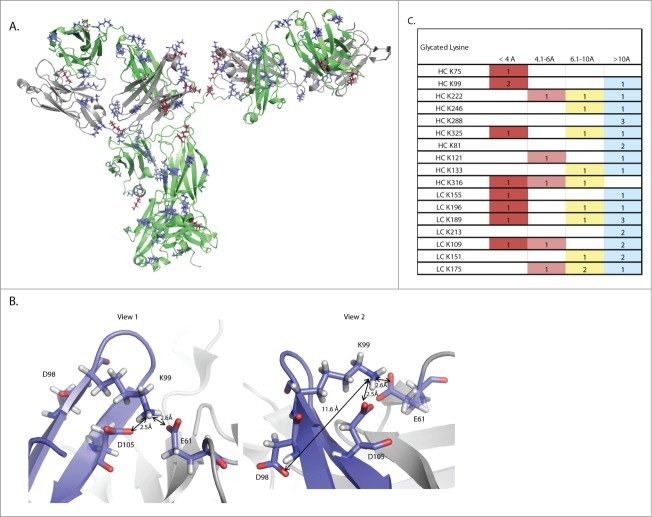
Figure 5.
Structural modeling of this mAb reveals a putative mechanism for mediation of non-enzymatic glycation. (A) A three dimensional ribbon model of the mAb. The heavy chain is shown in green while the light chain is shown in gray. Lysine residues are shown in blue, while glycated lysine residues are shown in burgundy. (B) Two views of the K99 glycation hot spot. Two residues on the heavy chain, D98 and D105, and one residue on the light chain, E61, are able to exert an effect on K99. The red coloring on D98, D105 and E61 shows the position of oxygen atoms in the carboxylic acid.

